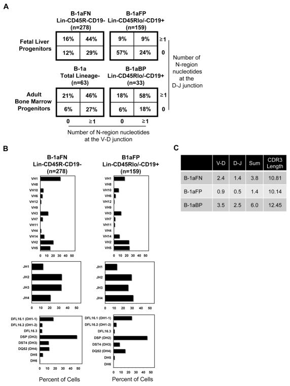
Figure 4. Immunoglobulin heavy chain sequence analysis of Lin-CD45R-CD19- fetal liver and Lin-CD45Rlo/-CD19+ bone marrow derived B-1a cells show abundant N-addition.
(A) N-region addition analysis at the D-J and V-D junctions in B-1a cells derived from Lin-CD45R-CD19- and Lin-CD45Rlo/-CD19+ fetal liver cells, Lin-CD45Rlo/-CD19+ bone marrow cells, and total Lin- bone marrow is shown. (B) V, D, and J gene segment usage in B-1a cells derived from Lin-CD45R-CD19- or Lin-CD45Rlo/-CD19+ fetal liver cells is displayed. (C) Average number of N-additions at the V-D, D-J, or sum of the two junctions is shown along with CDR3 length. Results are based on 3-5 independent experiments with sequences combined from each independent experiment.

