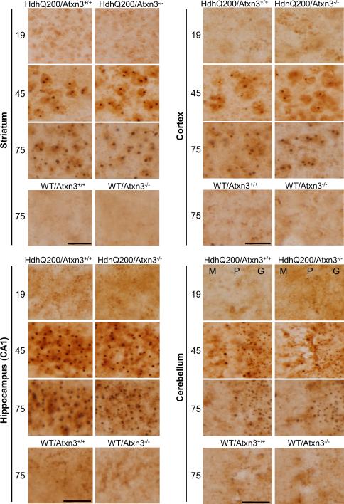
Fig.4. Loss of ataxin-3 does not alter inclusion body formation.
Immunohistochemistry with an N-terminal htt antibody was performed on brain sections from 19, 45 and 75 week old mice (n=3 per genotype). HdhQ200 mice show age-dependent accumulation of inclusion bodies in various brain regions (striatum, cortex, hippocampus and cerebellum). The size and density of inclusion bodies were similar in HdhQ200 mice with or without ataxin-3. Scale bars =20 μm. M (Molecular layer), P (Purkinje cell layer), G (Granule cell layer).

