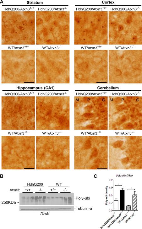
Fig.5. Deletion of ataxin-3 does not alter ubiquitination of htt inclusions, but increases total ubiquitinated proteins in wild type and HD mouse brain.
A) Anti-ubiquitin immunohistochemistry on brain sections from 75 week old HD mice (n=3 per genotype) show robust and abundant ubiquitinated inclusions in various brain regions (striatum, cortex, hippocampus and cerebellum), regardless of the presence or absence of ataxin-3. Scale bars =20 μm. M (Molecular layer), P (Purkinje cell layer), G (Granule cell layer).
B-C) Levels of total ubiquitinated proteins in forebrain of 75-week-old mice, determined by western blot, Show that loss of ataxin-3 significantly increased levels of ubiqutinated proteins in forebrain. Results are means ± SEM, * p<0.05. Blots were quantified with ImageJ (One-way ANOVA followed by post hoc test, n=3)

