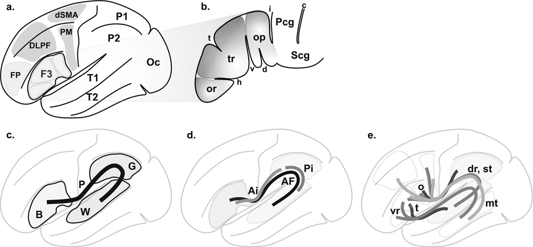
Figure 1. Language circuitry.
A. Overview of cortical language territories: inferior frontal gyrus (F3, or ventrolateral prefrontal cortex, or Broca’s area), superior temporal gyrus (T1), middle temporal gyrus (T2), premotor cortex (PM), dorsal supplementary motor area (dSMA), dorsolateral prefrontal cortex (DLPF) and inferior parietal gyrus (P2, or inferior parietal lobule). The frontopolar region (FP), the superior parietal gyrus (P1, or superior parietal lobule) and the occipital lobe (Oc) are added for further analysis. B. The Broca’s area (adapted from (Geschwind, 1970; Kirshner, 1995; Simmons-Mackie, 1997)): pars opercularis (op), pars triangularis (tr), pars orbitalis (or), horizontal (h) and vertical (v) segments of anterior lateral (or Sylvian) fissure, sulcus triangularis (t) and diagonalis (d). The central region is depicted (precentral gyrus, Pcg, subcentral gyrus, Scg, central sulcus, c and inferior precentral sulcus, i). C. Classical primary perisylvian circuitry (P) from Broca’s area (B) to Wernicke’s area (W); Geschwind’s territory (G) associated with AF, leading to conduction aphasia when lesioned (see(Catani et al., 2005)) is depicted. D. Circuitry according to Catani (Saur et al., 2008). The perisylvian network consists of a direct long pathway (AF) and an indirect pathway with anterior (Ai) and posterior (Pi) segments. E. Other circuitries: ventral (vr) and dorsal (dr, using AF) routes(Frey et al., 2008); opercular (o) and triangular (t) paths(Glasser & Rilling, 2008); superior (st, from operculo-premotor to T1 using AF) and middle (mt; from F3, PM and dorsolateral cortex to T2) temporal gyrus paths(Parker et al., 2005); Parker et al (Parker et al., 2005) results are not depicted because they overlap with the other information.

