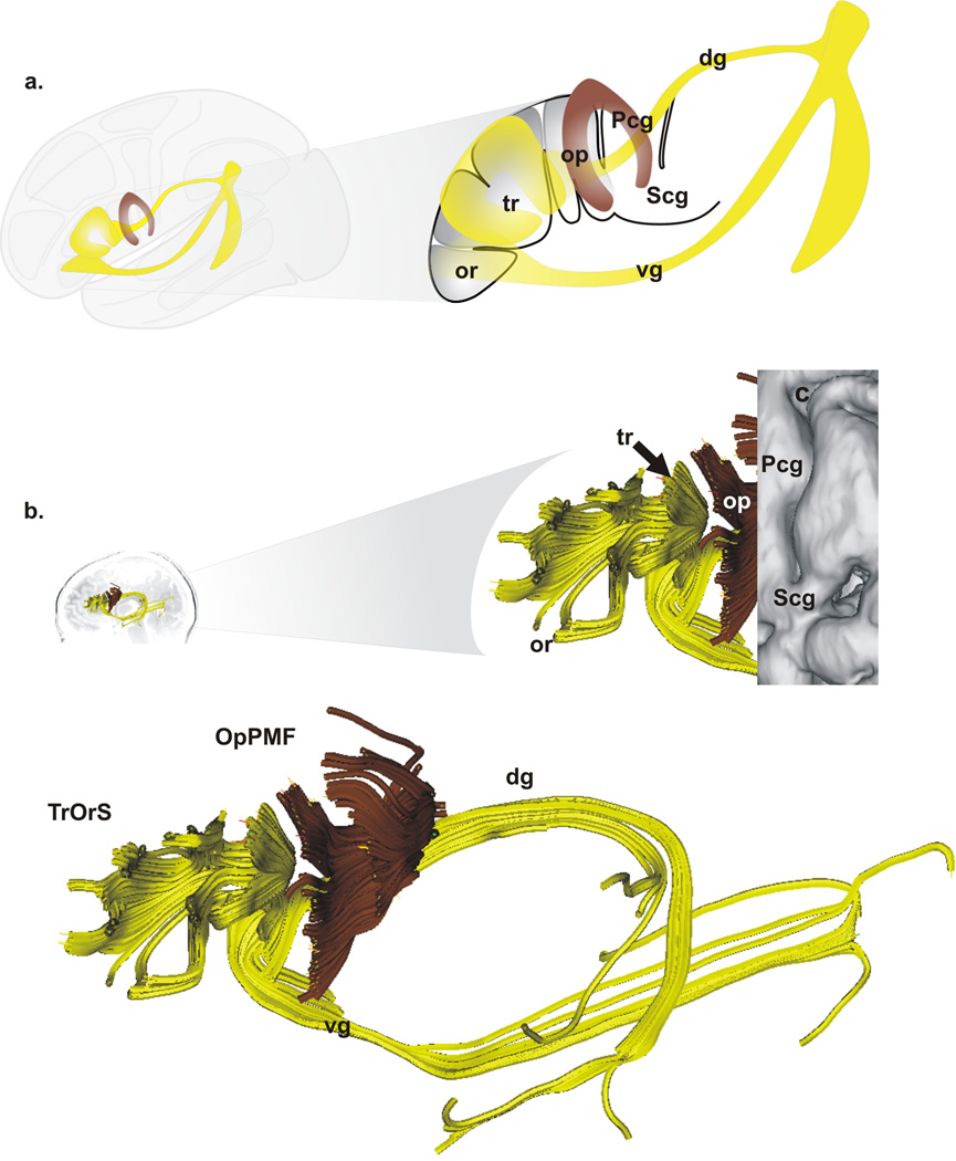
Figure 3. Fascicles of Broca’s area.
A. Schematic representation of the two fascicles identified within Broca’s area: the operculo-premotor fascicle (OpPMF, brown) and the triangulo-orbitaris system (TrOrS, yellow) as well as two groups of variable bundles (the dorsal, dg, and the ventral, vg, groups) linked with the TrOrS: left, overview; right, Broca’s area with the pars opercularis (op), the pars triangularis (tr), the pars orbitalis (or), the central region is depicted with the precentral gyrus (Pcg) and the subcentral gyrus (Pcg). B. Example of OpPMF and TrOrS (left hemisphere) within Broca’s area (overlay of the cortex, top right); bottom, details of the OpPMF and TrOrS fascicles.

