Figure 3.
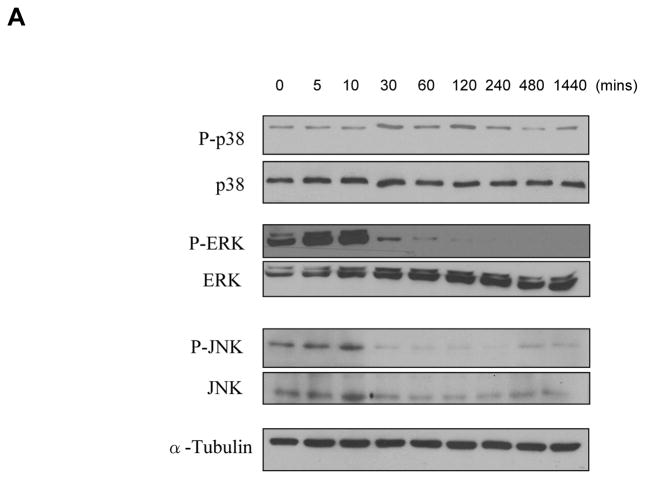
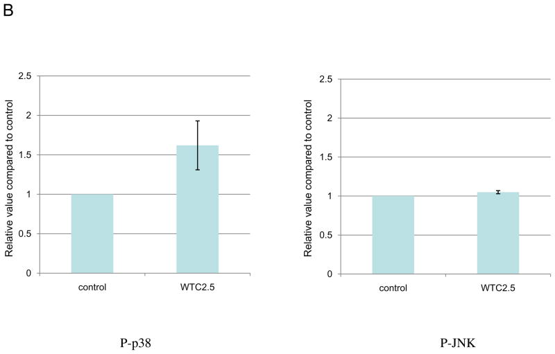
WTC2.5 effects on activation of ERK and p38 kinase pathways. (A) BEAS-2B cells were exposed to 100 μg WTC2.5/ml for varying time intervals, and the levels of phosphorylated form and total MAPK kinases (ERK, p38, and JNK) were assessed from Western blots using specific antibodies. The same membrane was blotted with α-tubulin antibody to verify protein loading in each lane. Because the same amounts of protein lysates from the same samples were used for the ERK, p38 and JNK blots, only one representative α-tubulin blot is shown here. (B) BEAS-2B cells were treated with 100 μg WTC2.5/ml; thereafter, JNK and p38 phosphorylation levels were measured at 10 and 60 min after start of exposure (as these represented the timepoints above where “maximal” phosphorylation appeared to have occurred). Western blot band intensities were quantified at each timepoint using an ImageJ program. Results (expressed as mean ± SD; n=2) shown represent relative intensity values from treated and untreated cells.


