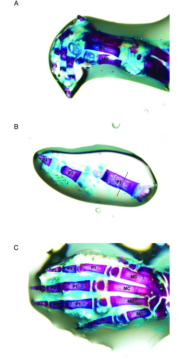
Figure 3.
Whole-mount bone preparations. The purple-stained tissue comprises bone, and the blue-staining tissue comprises cartilage. (A) Lateral view of PND 7 left forepaw. (B) PND 7 fifth (most lateral) digit of a hindpaw. The dotted line indicates the approximate site of amputation. (C) Dorsal view of PND 10 left forepaw. MC, metacarpal; P1, proximal phalanx; P2, intermediate phalanx; P3, terminal phalanx. Alizarin red S–Alcian blue double stain.

