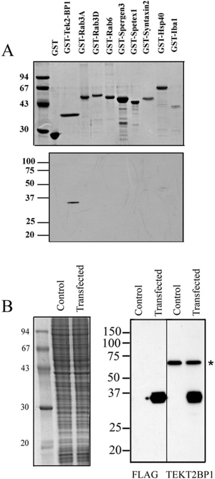
Figure 5.

Specificity of the anti-TEKT2BP1 antibody. (A) Recombinant GST-fused proteins were produced in E. coli and separated by SDS-PAGE. Separated proteins were stained with Coomassie brilliant blue (upper panel) or transferred to a PVDF membrane for immunoblotting with the purified antibody against TEKT2BP1 (lower panel). The antibody recognized a 35-kDa GST-TEKT2BP1 protein without any prominent cross-reactivity to other GST-fused proteins. Molecular mass standards are shown in kilo-Daltons (left). (B) COS7 cells expressing FLAG-tagged TEKT2BP1 were lysed, and the resultant proteins separated on SDS-PAGE. Separated proteins were either stained with Coomassie brilliant blue (left panel) or transferred to PVDF membranes (right panel) for immunoblot analysis using either the anti-FLAG antibody (FLAG) or the anti-TEKT2BP1 antibody (TEKT2BP1). Both antibodies recognized a protein migrating at 35 kDa, which was not detected in the control samples (non-transfected cells). The asterisk indicates a nonspecific protein band of COS7 cells recognized by the anti-TEKT2BP1 antibody. Molecular mass standards are shown in kilo-Daltons (left).
