Figure 2.
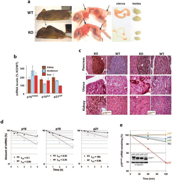
Aging-related phenotypes and upregulation of p16Ink4A, p19Arf and p21CIP expression in Auf1-/- mice. (a) Representative images of kyphosis (hunchback) (arrow), presence (top panel) and absence (bottom panel) of subcutaneous body fat (arrows), and atrophied gonads typically observed in 12-month-old Auf1-/- (KO) mice but not in WT littermates. Images shown represent the majority of KO animals surveyed (>60%, kyphosis; >90%, reduced body fat; >90%, gonadal atrophy). (b) Increased relative expression of p16Ink4a, p19Arf and p21CIP mRNAs in tissues and organs from 12-month-old KO mice compared to WT littermates, determined by qRT-PCR. (c) Immunohistochemical staining for p16Ink4a and p21CIP (brown, arrows) in organ sections from 12-month-old KO mice compared to WT littermates. Sections counter-stained by H&E. (d) Decay plots of p16Ink4a, p19Arf and p21CIP mRNAs in MEFs from G2 embryos. Transcription was blocked with actinomycin D, cells were collected at indicated time points, RNA was extracted and mRNA levels were quantified by qRT-PCR. Plots are the mean of three independent experiments with calculated half-lives shown. (e) Decay plots showing recovery of p16Ink4a mRNA destabilization by individual isoforms of AUF1 in KO MEFs. Plot shown is an average of three independent experiments.

