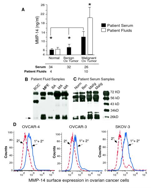
Figure 1. MMP-14 is a biomarker of malignant ovarian cancer.
A. Serum samples were collected from females with malignant ovarian cancer (n= 26), females with benign ovarian masses (n= 32) and normal females (n= 34) and MMP-14 cellular exodomain levels were measured by ELISA. Various fluids were collected from 14 patients with benign or malignant disease. MMP-14 were measured by ELISA in malignant ovarian ascites (n=5), malignant ovarian cyst (n=1), pleural effusion of ovarian cancer patients (n=4), benign ascites (n=3) and benign ovarian cyst (n=1) B. Columns mean, bars SE; * P<0.05, * * P<0.01. B and C. Gels showing cleaved MMP-14 fragments in benign and malignant patient fluids. Patient serum and fluids (BOC=benign ovarian cyst, BA=benign ascites, MA=malignant ovarian ascites, MPE =malignant pleural effusion) were diluted, mixed with citrate buffer and immunoblotted with MAB 918 in 1:1000 dilution. The cleaved MMP-14 fragment seen at 27 K Da is most representative of the ELISA data shown above D. Surface expression of MMP-14 was measured in ovarian cancer cell lines OVCAR4, OVCAR-3 and SKOV-3 by flow cytometry using a MMP-14 monoclonal antibody. The red dotted line represents secondary alone and the blue solid line represents primary plus secondary antibody.

