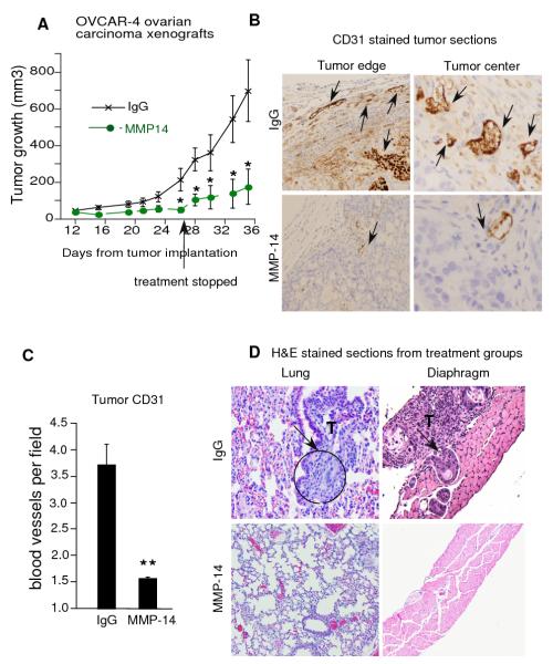
Figure 5. MMP-14 antibody inhibits tumor growth, angiogenesis, invasion and metastasis in mice.
A. OVCAR-4 ovarian cancer cells (3.5 million) were injected into the flanks of nude mice and the mice were treated with either IgG or MMP-14 antibody, i.p. 24 hours after implantation. Treatment was stopped from Day 27 onwards but tumors were monitored and measured by calipers every other day till day 35. B and C. Tumors were stained with CD31 antibody and blood vessels counted in sections from each tumor. Intense staining of individual endothelial cells was scored as a blood vessel in a blinded manner from paraffin embedded tissue sections. Vascular density was determined by counting 5-9 fields (160X magnification) from at least 5 tumors in each group. CD 31 stained blood vessels are demonstrated by black arrows in tumor edge and center. Columns mean, bars SE; * P<0.05, * * P<0.005. D. Female NCR Nu/Nu mice were injected i.p. with 1.5 million OVCAR-4 cells and randomized into treatment groups of IgG (n=5) or MMP-14 Ab (n=4, 2.5 mg/kg/day, twice a week, i.p.), starting on day 2. Both groups received docetaxel (5 mg/kg/week, i.p.) starting on day 21 and a repeat dose after 7 days. Mice were euthanized and diaphragm, peritoneal, and thoracic organs were harvested, stained with hematoxylin & eosin and analyzed by two independent pathologists blinded to the treatment groups. Representative sections from lung and diaphragm are shown and tumor areas are marked by arrows.

