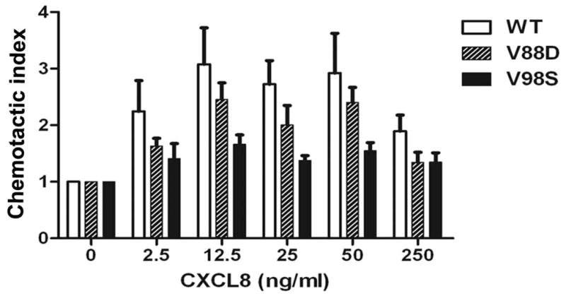
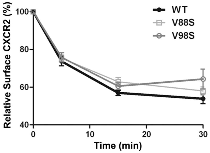
Figure 6. Over-expression of the dominant negative AP2-σ2 inhibits CXCR2-mediated chemotaxis without significantly affecting the internalization of CXCR2.


A) WT and dominant negative V88D and V98S AP2-σ2 constructs were transiently over-expressed in HEK-293-CXCR2 cells and chemotaxis assays were performed in a modified Boyden chamber as described in ‘Methods’. Experiments were repeated 3 times with triplicates in each treatment. ANOVA: WT vs. V88D, p = 0.03; WT vs. V98S, p < 0.001. B) CXCR2 internalization assay was performed with FACS analysis as described in ‘Methods’. The geometric mean of the fluorescence intensity was used to calculate the percentage of cell surface CXCR2. ANOVA: WT vs. V88D, p= 0.0262; WT vs. V98S, p = 0.0279. Bonferroni post-hoc tests – WT vs. V98S at 30 min (p < 0.05).
