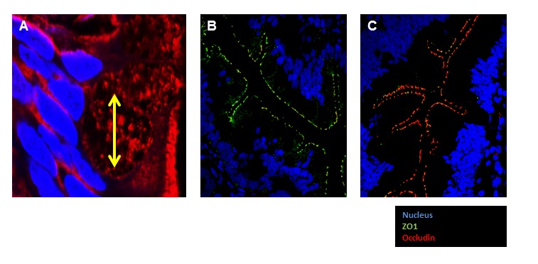
Figure 2.

Fluorescent immunohistochemical staining of jejunum tight junction proteins. (A). Goblet cell diameter is marked in yellow. Nucleus: Blue Dapi, Cell Membrane: Orange mask. Image was obtained at 100X oil immersion confocal microscope. (B and C) IHC demonstrating the expression of Tight junction proteins, occluding (red) and ZO1 (Green) in small intestinal tissue (60X).
