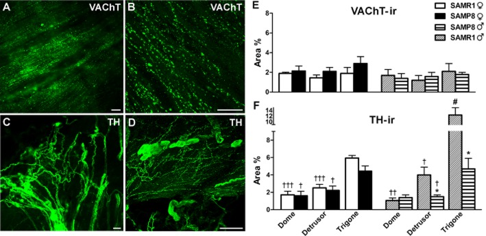
Figure 4.

VAChT-and TH-ir in SAMR1 and SAMP8 bladders. (A) Abundant VAChT-ir cholinergic nerves are evident in the male SAMP8 detrusor muscle. (B) Confocal image showing z-stacks of a 2 μm section of the trigone from a male SAMP8 bladder in which VAChT-ir nerves can be observed running parallel to the muscle bundles. (C) A high density of TH-ir adrenergic nerve trunks can be seen in the trigone from a male SAMR1 bladder. (D) Confocal image showing z-stacks of a 2 μm section of the trigone muscle layer from a female SAMR1 bladder in which abundant TH-ir nerves can be observed. Note the presence of TH-ir ganglion cells. Scale bar = 25 μm. Quantification of VAChT-ir (E) and TH-ir (F) in SAMR1 and SAMP8 bladders. Measurements were acquired independently in different regions of the bladder muscle layer (dome, detrusor and trigone). Hand-drawn fields of whole-mount preparations were selected and the area above the intensity threshold was measured (Area %). The values represent the mean ± SE of three to five different fields from four to six different animals: *P < 0.05 versus SAMR1; #P < 0.05 versus female, and †P < 0.05, ††P < 0.01 and †††P < 0.001 versus the trigone region (anova followed by Student's t-test).
