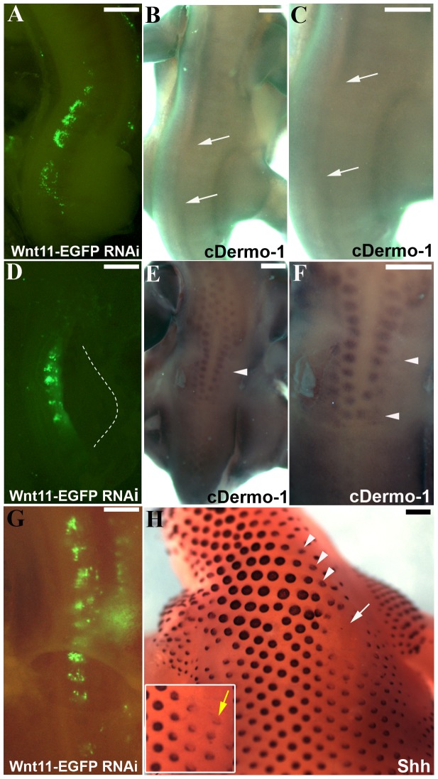
Figure 4. A series of 9 photos of a chicken embryo section electroporated with a Wnt11 RNAi construct containing EGFP at the DML level, and than reincubated for 24 h.
Each photo shows movement of cells in 1 h time interval. In contrast to the control experiment (Figure 3), the electroporated cells (green fluorescent) remain restricted to the DML or migrate into the myotome. No EGFP positive cells migrate towards the subectodermal space located above the neural tube or into the immediate neigbourhood. The absence of Wnt11 in the DML thus results in a compromised EMT of the cells, which can only enter the myotome and no longer populate the subectodermal space above the neural tube. The red outline indicates the DML. Scale bar: 100 μm.

