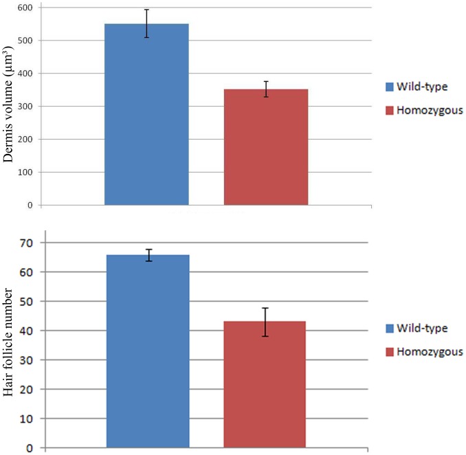
Figure 8. Dorsal dermis is decreased in dermis thickness and number of hair follicles in the Wnt11 knock-out mice.
A, C, E. Homozygous Wnt11 knock-out embryos. B, D, F. Wild-type embryos. Embryo in A represents a homozygous Wnt11 knock-out embryo of embryonic day E 14.5. In the dorsal mid-line region of this embryo a large area devoid of hair placodes is detectable. The white square shows a decrease number of hair placodes in comparison to the WT embryo of similar developmental stage (B). Section through the embryo in A shows that the cDermo-1 signal is diminished with corresponding decrease in dermal thickness in the mutant (black arrow in C) as compared with a similar section of the same magnification of a WT embryo (black arrow in D). Hematoxylin-Eosin staining in paraffin sections of knock-out mice of E 18.5 shows a remarkable decrease in dermis thickness (45%), as well as a reduced number of hair follicles (35%). (E) In comparison with a Hematoxylin-Eosin treated section of a WT mouse of the same developmental stage (F). Double-headed arrows in E and F show the remarkable difference in dermis thickness between the mutant mouse embryo as compared to the normal thickness of dermis in a wild-type embryo (photographs were taken at the same magnification). The sections were taken at the trunk regions of the mice. The analysis was restricted to the same anatomical area of the dorsal skin for all embryos investigated. Scale bar: 100 μm.

