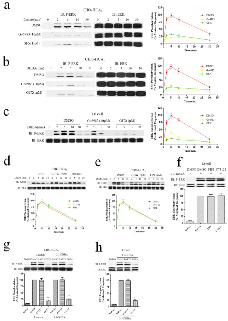
Figure 4. Effects of PKC, PLC, PLD and calcium on HCA1-stimulated phosphorylation of ERK1/2.
Serum-starved CHO-HCA1 cells (a and b) or L6 cells (c) were pretreated with DMSO or 10 μM Go6983 or 1 μM GF109203X (GFX) for 1 h, and then stimulated with 10 mM L-Lactate (a) or 300 μM 3,5-DHBA (b) for CHO-HCA1 or 3 mM 3,5-DHBA for L6 cells (c) for the indicated time periods. Serum-starved CHO-HCA1 cells (d and e) or L6 cells (f) were pretreated with DMSO or 20 μM U73122 or 1 μM FIPI for 1 h, and then stimulated with 10 mM L-Lactate (d) or 300 μM 3,5-DHBA (e) for CHO-HCA1 cells for indicated time periods, and 3 mM 3,5-DHBA for L6 cells (f) for 5 min. Serum-starved CHO-HCA1 cells (g) or L6 cells (h) were cultured in serum-free DMEM/F12 or DMEM media with or without EGTA (5 mM) or BAPTA-AM (50 μM) for 1 h, cells were then stimulated with 10 mM L-Lactate or 300 μM 3,5-DHBA (g) for CHO-HCA1 and 3 mM 3,5-DHBA for L6 cells (h) for 5 min. The data shown are representative of at least three independent experiments. Error bars, S.E. for three replicates. Data were analyzed by using the Student’s t test (***p<0.001). IB, immunoblot; P-ERK, phospho-ERK.

