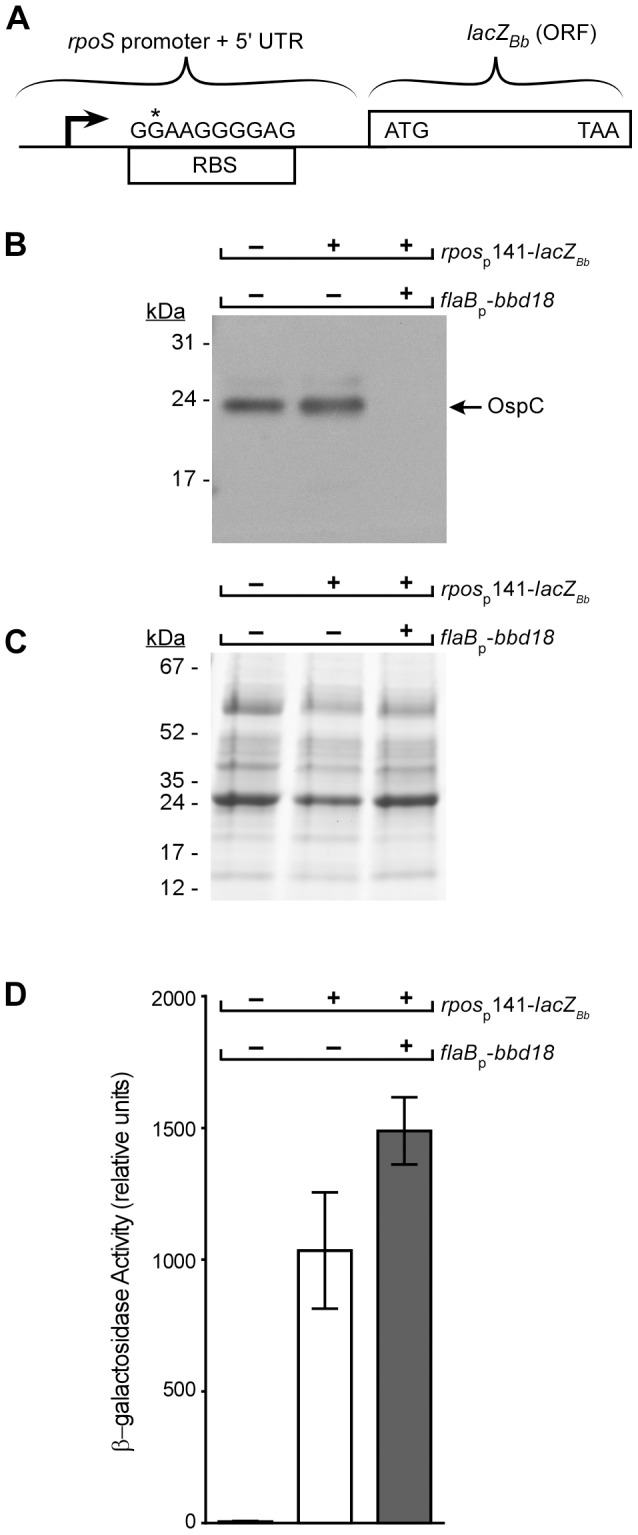
Figure 5. Analysis of BBD18 repression of an rpoS promoter lacZBb transcriptional fusion.

(A) A schematic diagram of the transcriptional fusion of the rpoS promoter and 5’ untranslated region (UTR) fused directly to the lacZ Bb open reading frame (ORF). The position of the rpoS transcriptional start site [58], [63] is indicated by a filled arrowhead. The Shine-Dalgarno sequence (RBS), and the translational start site of β-galactosidase (lacZBb), indicated by the ATG, are also shown. Regions corresponding to the rpoS promoter and 5’ UTR (141bp) or the lacZ Bb ORF are indicated with brackets. The "G" in the RBS marked with an asterisk is identified because the reporter construct harbored an A->G mutation relative to the published sequence at that position. Cell lysates from strains B31-S9 (wt), B31-S9/pBSV2G-rpoSp141-lacZBb (wt/rpoSp-lacZBb), and B31-S9/pBSV2G- rpoSp141-lacZBb/pBSV28-flaBp-bbd18 (wt/rpoSp-lacZBb/flaBp-bbd18) were grown under rpoS-inducing conditions and analyzed with OspC antisera (B) or stained with Coomassie blue (C) to demonstrate equivalent protein loads in each lane. The positions of molecular mass standards are shown on the left in kiloDaltons (kDa). (D) β-galactosidase activity in cell lysates from strains B31-S9 (wt), B31-S9/pBSV2G-rpoSp141-lacZBb (wt/rpoSp-lacZBb), and B31-S9/pBSV2G- rpoSp141-lacZBb/pBSV28-flaBp-bbd18 (wt/rpoSp-lacZBb/flaBp-bbd18) grown under rpoS-inducing conditions.
