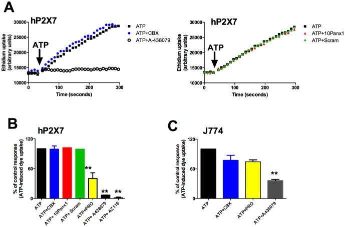
Figure 1. Effect of pannexin-1 inhibitors on ATP-induced dye uptake in HEK-293 cells expressing human P2X7 and J774 macrophages.
(A) Ethidium+ uptake was induced in HEK-hP2X7 expressing cells by the addition of 1 mM ATP (denoted by the arrow) in low divalent physiological solution. Control ethidium uptake to ATP is shown in black squares. Cells were pre-incubated with inhibitors for 10 minutes at 37°C before ATP addition. In the top panel ethidium uptake in the presence of 10 μM carbenoxolone (CBX) is shown in blue circles and in the presence of 10 μM A438079 in black open circles. In the lower panel ethidium uptakes in the presence of the pannexin-1 peptide (10Panx1) or scrambled 10Panx1 peptide are shown in red triangles or green diamonds respectively. Cellular fluorescence was measured using a fluorescent plate reader over 300 seconds. (B) Bar chart displaying mean data from 2–5 separate experiments using inhibitors carbenoxolone (CBX), 10Panx1 peptide or scrambled 10panx1 peptide (100 μM), A-438079 and AZ11645373. Data was calculated at 300 second timepoint as baseline corrected endpoint data and normalised to the ATP control. (C) ATP (3.4 mM) induced ethidium uptake was measured in J774 macrophages. CBX (50 μM), PRO (2.5 mM) and A438079 (10 μM) significantly reduced the response. Error bars represent S.E.M, ** P<0.05 calculated by one-way ANOVA (Dunnett's post-test).

