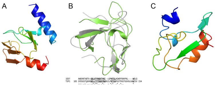
Figure 3. Folds of non β-helix subdomains of TSP1.
(A) Overall fold of the D1 subdomain. The chain is colored progressively from blue (N-terminus) to red (C-terminus). (B) Structure homology between subdomain D2 (green) and the chitin binding domain of Chitinase from Bacillus circulans (gray). The structure based sequence alignment shows the well-superposed residues in bold underlined letters. Invariant residues are marked by *. (C) Overall fold of the D3-D4 linker region. The chain is colored progressively from blue (N-terminus) to red (C-terminus).

