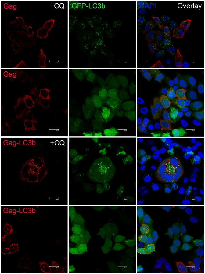
Figure 2. Functionally targeted SIV Gag protein to autophagosomes using confocal microscopy.
HeLa-GFP-LC3 cell line stably expressing GFP-LC3 protein (green fluorescence) were transfected with pVAX-SIVgag plasmid (top) or pVAX-SIVgag-LC3 plasmid (bottom) with or without chloroquine (CQ) treatment, and then stained with anti-SIVgag and Cy3-labeled goat anti-mouse IgG as secondary antibodies (red fluorescence); the nuclei were stained with DAPI (blue fluorescence). The scale bar represents 20 μm. Representative cells are shown from one experiment out of three total experiments.

