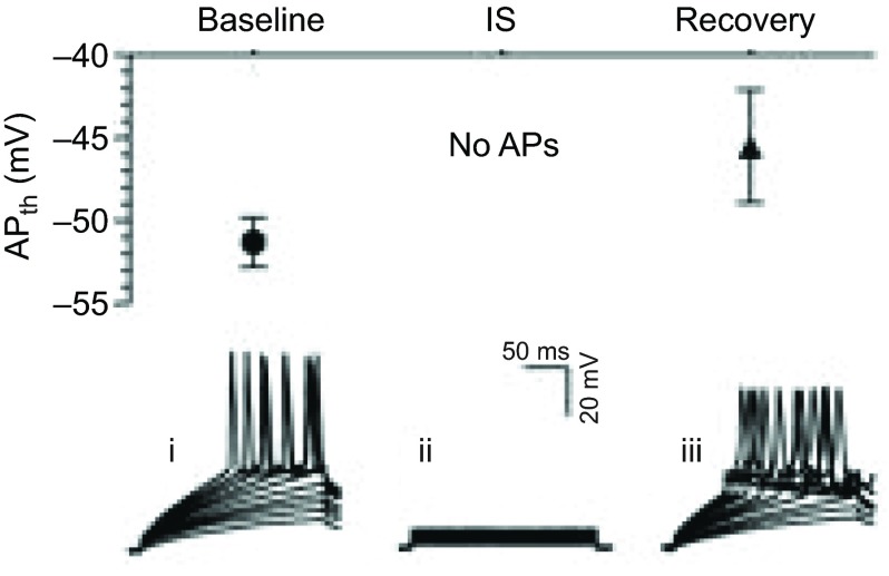
Fig. 2.
Cortical neuronal membrane potential (mV) treated with an ischemic mimicking solution depolarizes to EGABA. Top panel: summary of action potential (AP) threshold (APth) from stimulated neurons treated as indicated. APs could not be elicited during IS (ischemic solution) treatment. Bottom panel: sample recordings of evoked APs recorded during baseline control (i), IS perfusion (ii) and normoxic reperfusion (iii). [Adapted from Pamenter et al. (Pamenter et al., 2012); reprinted with permission from Macmillan Publishers Ltd.]

