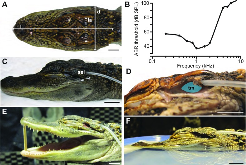
Fig. 11.
Photographs of the preparation setup and audiogram. (A) Illustration of the morphological measurements reported in Table 1: head diameter at the widest point (d), length of head (l) and the interaural distance (ia) measured as the distance between ear ridges. (B) In-air ABR audiogram based on data from Higgs et al. (Higgs et al., 2002), showing low frequency hearing specialization. (C) For auditory space measurements, the earlid was trimmed to achieve a naturalistic position. The microphone probe tube was threaded through the superior earlid (sel) and secured with liquid adhesive. (D) The earlid has been cut away to show the position of the probe with respect to the tympanic membrane (tm). (E) Image of the mouth-open condition (see Materials and methods for details). (F) For the water surface condition the head was positioned in a shallow plastic container filled with water to mimic a naturalistic floating position (see Materials and methods for details). Scales bars, 10 mm.

