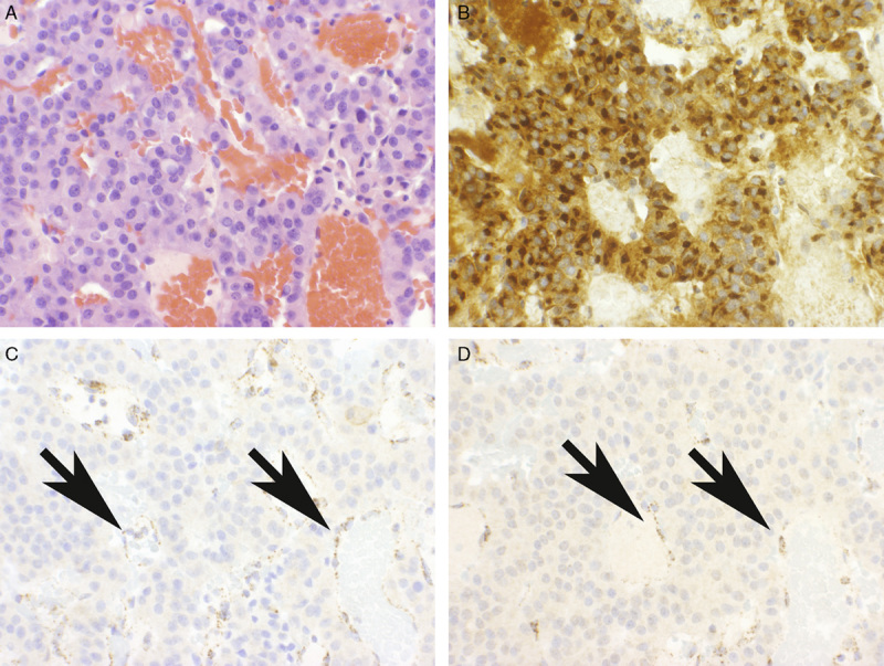
FIGURE 2.

Pituitary adenoma demonstrating loss of staining for SDHB and SDHA. The neoplastic area (A), which demonstrates uniform strong prolactin expression (B), demonstrates completely absent staining for both SDHA (C) and SDHB (D). Note: The non-neoplastic endothelial cells (arrows) demonstrate strong positive staining for both SDHA and SDHB and serve as a positive internal control (A, H&E; B, Prolactin; C and D, IHC).
