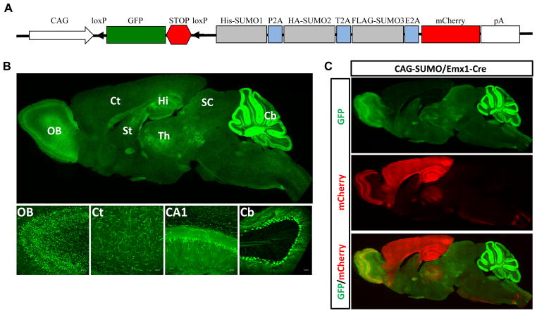
Figure 1.
Generation of CAG-SUMO transgenic mice. A, Scheme of the transgene construct. The transgene consists of the CMV early enhancer/chicken β-actin promoter (CAG), a fragment containing GFP and a transcriptional/translational STOP cassette (STOP) flanked by loxP sites, 3 tagged SUMOs linked by 2A sequences, mCherry, and a polyadenylation signal (pA). B, Expression patterns of native GFP fluorescence in a CAG-SUMO line 10 mouse brain. A sagittal brain section indicated widespread GFP expression (upper panel). Lower panel shows confocal images of different areas of the brain. C, A sagittal brain section of a double transgenic CAG-SUMO/Emx1-Cre mouse showing the mCherry expression, indicative of expression of tagged SUMOs, restricted to forebrain regions. OB, olfactory bulb; Ct, cortex; Hi, hippocampus; CA1, hippocampal CA1 subfield; St, striatum; Th, thalamus; SC, superior colliculus; Cb, cerebellum. Scale bars: 50 μm.

