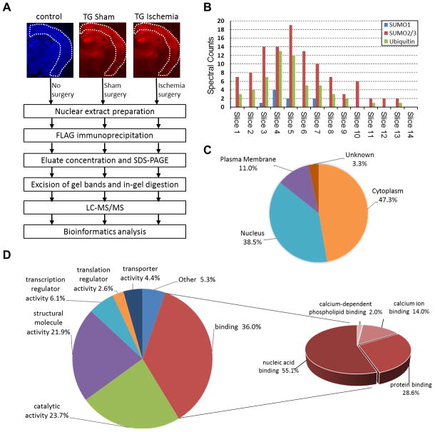
Figure 4.
Proteomics analysis of SUMO3-conjugated proteins in post-ischemic mouse brains. A, Overview of the workflow to identify FLAG-SUMO3-conjugates in the post-ischemic cerebral cortex. Coronal brain sections of Emx1Cre/+ (control; DAPI staining, blue) and CAG-SUMO/Emx1-Cre (TG; mCherry fluorescence, red) mice were shown to indicate cortical regions used in the study. B, Distribution of total spectral counts for SUMO1, SUMO2/3, and ubiquitin for each gel slice of ischemia samples from high (Slice 1) to low (Slice 14) molecular weights. C–D, Subcellular localization (C) and molecular functions (D) of the 91 putative SUMO3 substrates with upregulated SUMO3 conjugation state after ischemia were grouped by the PANTHER program.

