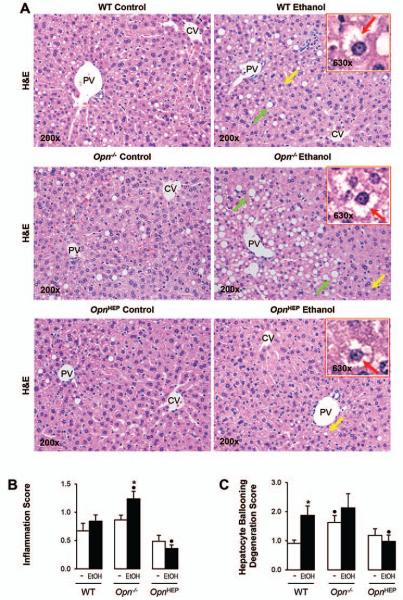
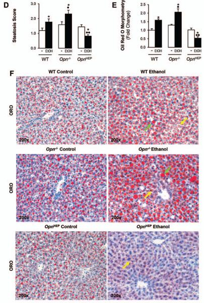
Figure 3. Natural induction plus overexpression of OPN in hepatocytes protects from alcohol-induced steatosis whereas natural induction of OPN does not suffice to confer full protection.
WT, Opn−/− and OpnHEP Tg mice were fed 7 wks either the control or the alcohol Lieber-DeCarli diet. H&E staining shows less inflammation, hepatocyte ballooning degeneration (red arrows on the insets), micro- (yellow arrows) and macrovesicular (green arrows) steatosis in ethanol-treated OpnHEP Tg followed by WT and by Opn−/− mice. PV: portal vein. CV: central vein (A). The scores for inflammation (B), hepatocyte ballooning degeneration (C) and steatosis (D) are lower in ethanol-treated OpnHEP Tg followed by WT and Opn−/− mice. Morphometric analysis (E) and oil red O staining (F). n=10/group; *p<0.05 for ethanol vs control; •p<0.05 and ••p<0.01 for any genotype vs WT mice.


