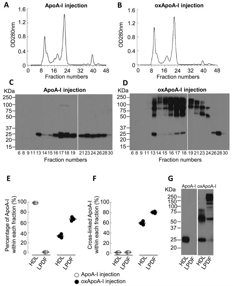
Figure 1. Distribution of injected human native and oxidized ApoA-I in the plasma of ApoE−/− mice.
ApoE−/− mice (16 weeks on Western diet) were injected s.c. with 15 mg of either native or oxidized (oxApoA-I) human ApoA-I. Blood was collected at 8 hours after the injections. A-B) Fast performance liquid chromatography (FPLC) on a Superdex 200 column was performed on pooled plasma (100 μl) from 3 mice. C-D) Western blot analysis of human ApoA-I in numbered FPLC fractions probed with anti-total human ApoA-I monoclonal antibody (mAb 10G1.5). E-G) HDL-containing lipoprotein fraction and lipoprotein deficient fraction (LPDF) were isolated from 40 μl of plasma after sequential buoyant density ultracentrifugation. Human ApoA-I levels were quantified by Western blot analysis using mAb 10G1.5. E) Percentage of injected ApoA-I within HDL versus LPDF, F) percentage of cross-linked ApoA-I within HDL versus LPDF and G) illustrative Western blot analyses probed with mAb 10G1.5 of the distribution of injected human native and oxidized ApoA-I in the plasma compartments.

