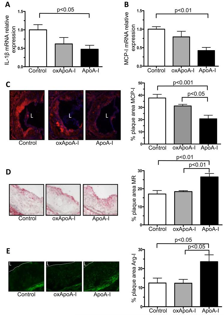
Figure 5. Inflammatory state of the plaque: changes in M1 and M2 macrophage markers.
A) IL1-β and B) MCP-I mRNA expression of laser-captured CD68+ aortic root plaque cells measured by qRT-PCR; minimum of 8 mice in each group, and immunohistochemistry for C) MCP-I, D) mannose receptor 1 (MR) and E) arginase-I (Arg-I) in aortic root plaques from ApoE −/− mice (16 weeks on Western diet) after s.c. injection (every other day) of native ApoA-I, oxidized ApoA-I (oxApoA-I) or carrier (control) over 1 week; magnification 10×, arginase-I 20×; minimum of 5 mice in each group; Data are shown as mean ± SEM; L = lumen.

