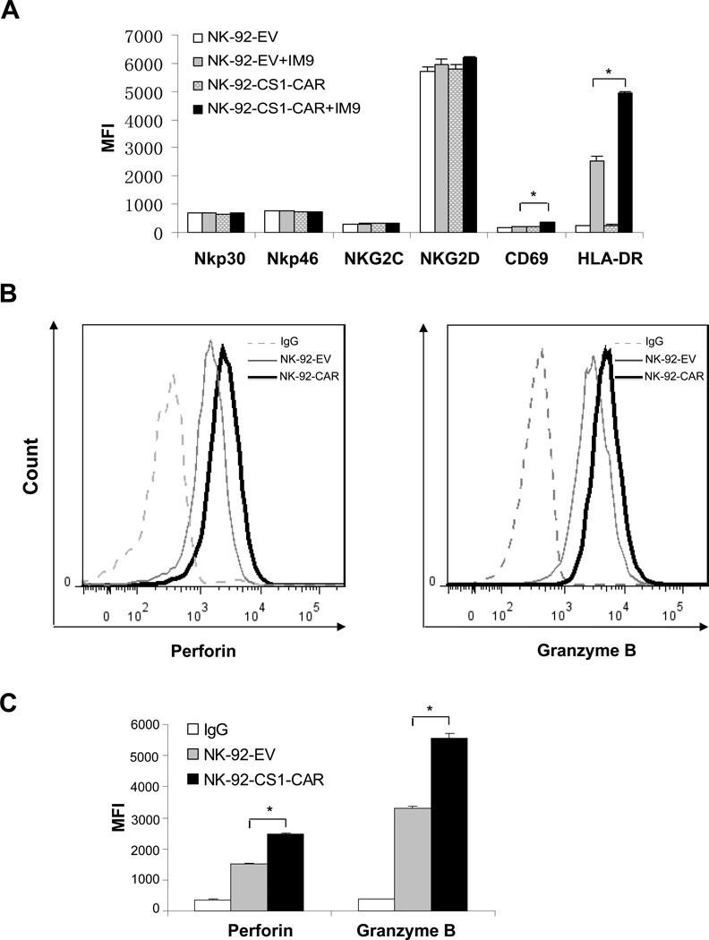
Figure 5. Phenotypic characterization of CS1-CAR modified NK cells.
A) Mock- or CS1-CAR-transduced NK-92 cells (NK-92-EV and NK-92-CS1-CAR, respectively) were either cultured alone, or cultured with IM9 MM cells for 4 h. Surface expression of NKp30, NKp46, NKG2C, NKG2D, CD69 and HLA-DR was assessed by flow cytometry following staining with corresponding monoclonal antibodies (mAbs), and the mean fluorescence intensity (MFI) was recorded. * indicates P < 0.05. B) NK-92-EV and NK-92-CS1-CAR cells were permeabilized for intracellular staining with mAb specific for perforin or granzyme B, and analyzed by flow cytometry. The dotted line represents staining the NK-92-EV control cells with CS1 mAb, solid heavy line for NK-92-CS1-CAR cells with CS1 mAb, and the dashed line for the NK-92-EV control cells with isotype-matched control antibodies. C) Depicted MFI for histograms shown in (B).* indicates P < 0.05.

