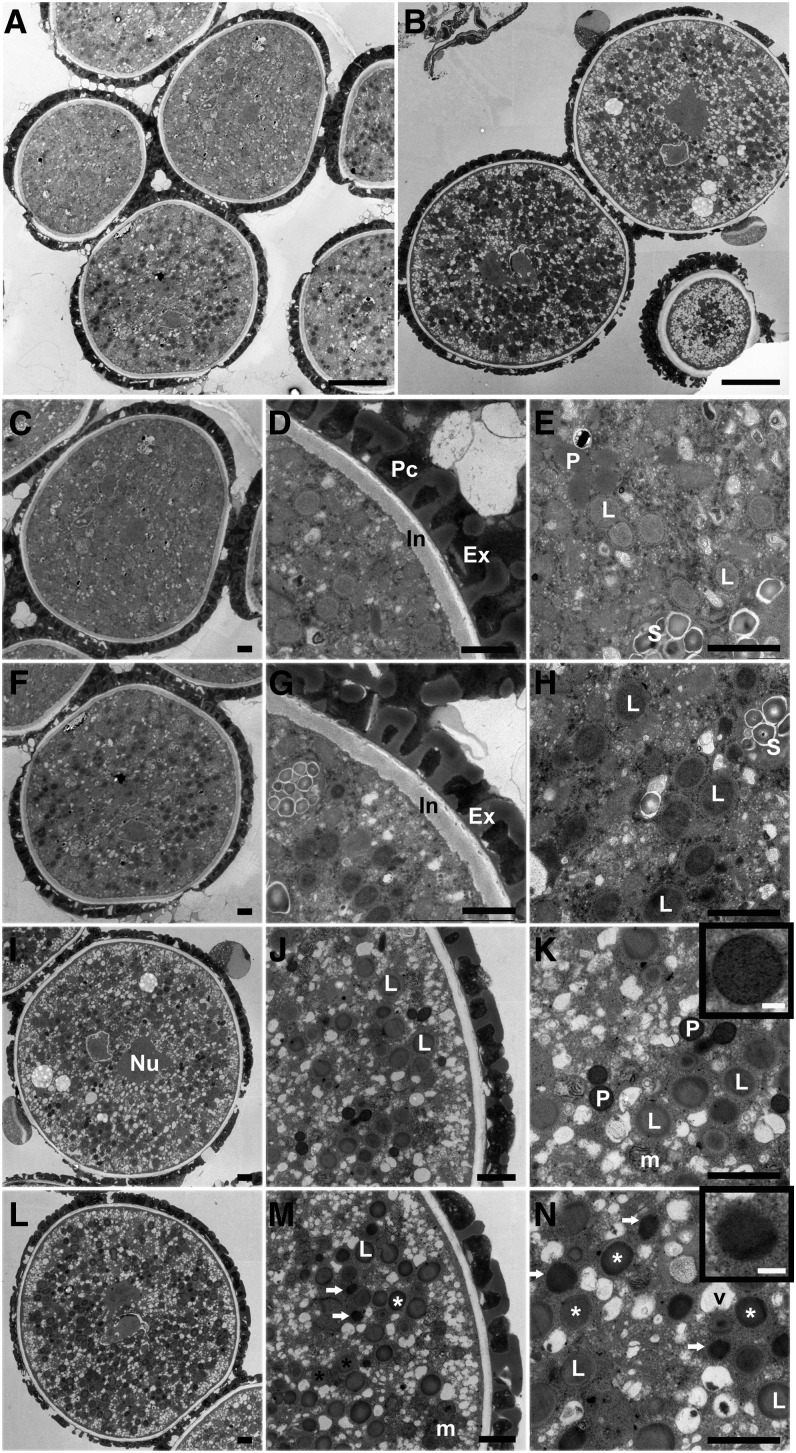
Figure 2.
TEM Analysis of dau Pollen Compared with the Wild Type.
(A) dau/DAU qrt/qrt tricellular pollen grains in undehisced anthers.
(B) dau/DAU qrt/qrt mature grains, stained with DAB.
(C) A wild-type tricellular pollen grain in undehisced anthers.
(D) A portion of (C) showing wild-type pollen cell wall.
(E) A magnification of (C) showing peroxisomes, lipid bodies, and the starch granules as indicated.
(F) A dau mature pollen grain in undehisced anthers.
(G) A portion of (F) showing dau mutant pollen cell wall.
(H) A magnification of (F) showing darkly stained lipid bodies.
(I) to (N) TEM micrographs of mature pollen released from dehiscent anthers with DAB staining.
(I) A wild-type mature pollen grain. The nucleus of one sperm cell is indicated.
(J) A magnified region of (I) showing gray lipid bodies.
(K) Detail of (I) showing heavily stained peroxisomes with clear boundary (inset), lipid bodies, and mitochondria. Mitochondrial cristae were also stained by DAB.
(L) An overview of a dau mature pollen grain.
(M) A magnified region of (L). Note DAB-stained peroxisome-like structure (arrow) and two types of lipid body (star).
(N) A magnification of (L) showing darkly stained peroxisome-like structures (arrow and inset) and stained (white star) and nonstained (dark star) lipid bodies.
Ex, exine; In, intine; L, lipid body; Nu, nucleus; m, mitochondria; P, peroxisome; Pc, pollen coat; S, starch granule. Bars = 5 µm in (A) and (B), 1 µm in (C) to (N), and 0.1 µm in the insets of (K) and (N).

