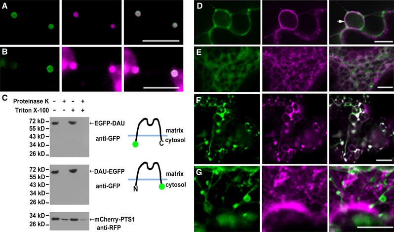
Figure 3.
Topology of DAU and Localization of DAU Truncated Protein.
(A) Coexpression of EGFP-DAU and the peroxisomal marker mCherry-PTS1 in tobacco leaves, showing peroxisomal membrane-localized EGFP-DAU and peroxisomal matrix-localized mCherry-PTS1.
(B) Coexpression of DAU-EGFP and peroxisomal marker mCherry-PTS1 in tobacco leaves, showing that DAU-EGFP is targeted to the peroxisomal membrane and mCherry-PTS1 is localized in the peroxisomal matrix and cytosol.
(C) Determination of DAU topology through the proteinase protection assay. Both the N terminus and C terminus of DAU face the cytosol. Peroxisomes from tobacco leaves expressing EGFP-DAU, DAU-EGFP, and mCherry-PTS1 were subjected to proteinase K treatment in the presence or absence of Triton X-100. Treated samples were subjected to SDS-PAGE and immunoblot analysis.
(D) Coexpression of DAU-EGFP and the ER marker mCherry-HDEL in perinuclear ER in tobacco leaves. Arrow indicates the nuclear membrane.
(E) Coexpression of EGFP-DAU(1-115) and the ER marker mCherry-HDEL in tobacco leaves.
(F) Coexpression of DAU(267-333)-EGFP and mCherry-PTS1 in tobacco leaves. Note the formation of tubular peroxisomes.
(G) Coexpression of DAU(267-333)-EGFP and mCherry-HDEL in tobacco leaves. Bars = 10 µm.

