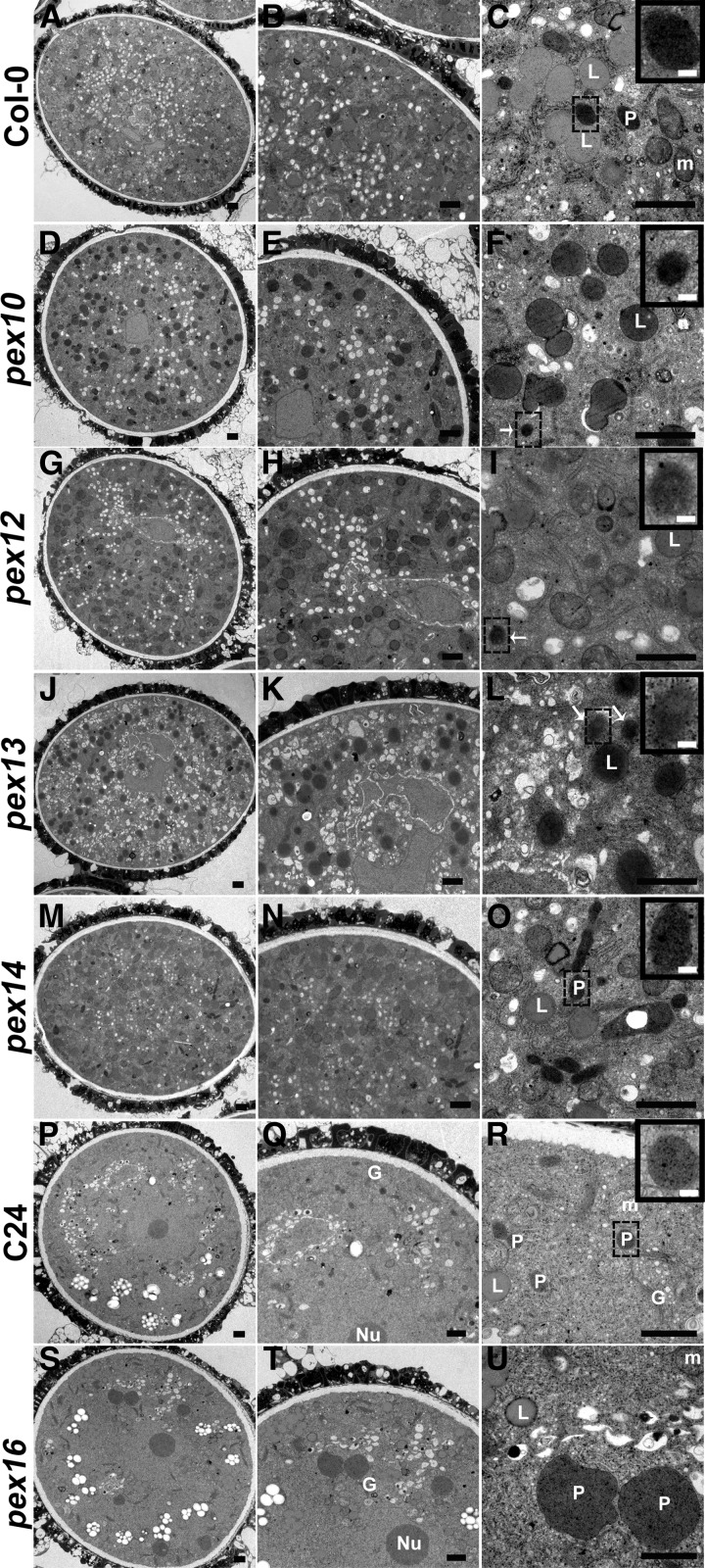
Figure 9.
TEM of pex Tricellular Pollen Compared with Wild-Type Pollen.
(A) to (C) Observation of a wild-type pollen grain, showing mitochondria, lipid bodies, and heavily stained peroxisomes with a clear boundary (inset). The boxed region is enlarged as the inset.
(D) to (L) TEM micrographs of pex10 ([D] to [F]), pex12 ([G] to [I]), and pex13 ([J] to [L]) pollen. Note darkly stained peroxisome-like structures (white arrow and insets) and heavily stained lipid bodies. Insets depict enlarged views of boxed regions.
(M) to (O) Observation of a pex14 pollen grain, showing heavily stained peroxisomes with a clear boundary (inset). The inset is an enlarged view of the boxed region.
(P) to (R) Observation of a C24 wild-type pollen grain, showing mitochondria, lipid bodies, and heavily stained peroxisomes with a clear boundary (inset). The boxed region is enlarged as the inset.
(S) to (U) TEM micrographs of pex16 pollen, showing huge peroxisomes with an irregular boundary.
L, lipid body; m, mitochondrion; P, peroxisome, G, Golgi apparatus. Bars = 1 µm in (A) to (U) and 0.1 µm in the insets of (C), (F), (I), (L), (O), and (R).

