Figure 10.
Association of Mitochondria with Autophagic Bodies during Leaf Senescence.
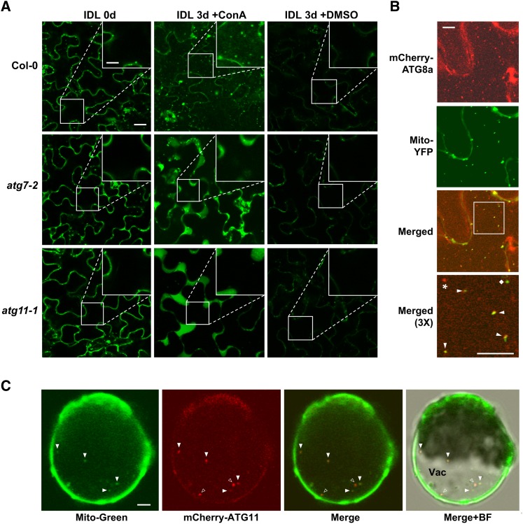
(A) Accumulation of Mito-CFP in vacuolar puncta during dark-induced leaf senescence (IDL) by a process that requires the ATG system. Four-week-old wild-type, atg7-2, and atg11-1 plants expressing Mito-CFP were subjected to IDL for 3 d followed by a 20-h incubation with 1 μM ConA or DMSO before confocal fluorescence microscopy of epidermal cells from the third and fourth rosette leaves. Bars = 10 μm and 3.4 μm in the inset. Insets show 3× magnifications of the vacuole.
(B) Colocalization of Mito-YFP with the autophagic membrane marker mCherry-ATG8a in autophagic bodies. Wild-type plants expressing both reporters were subjected to IDL senescence as in (A). The vacuolar region of leaf epidermal cells was imaged by confocal fluorescence microscopy. Bar = 10 μm. A 3× magnification of the merged signals (outlined by the white box) is included to confirm colocalization of the two proteins in autophagic bodies (arrowheads). A free Mito-YFP–labeled mitochondrion and an autophagic body not containing mitochondria are indicated with the diamond and the star, respectively.
(C) Colocalization of the mitochondrial stain MitoTracker Green FM with mCherry-ATG11. Arabidopsis leaf protoplasts stably expressing mCherry-ATG11 were treated for 30 min with MitoTracker Green FM, washed twice, and then incubated for 24 h with ConA in the absence of Suc before confocal fluorescence microscopy. Closed arrowheads indicate vacuolar puncta containing both fluorescent markers; open arrowheads are puncta preferentially containing mCherry-ATG11. BF, bright field; Vac, vacuole. Bar = 5 μm.

