Figure 3.
Interaction of ATG101 with Other Subunits of the ATG1/13 Kinase Complex.
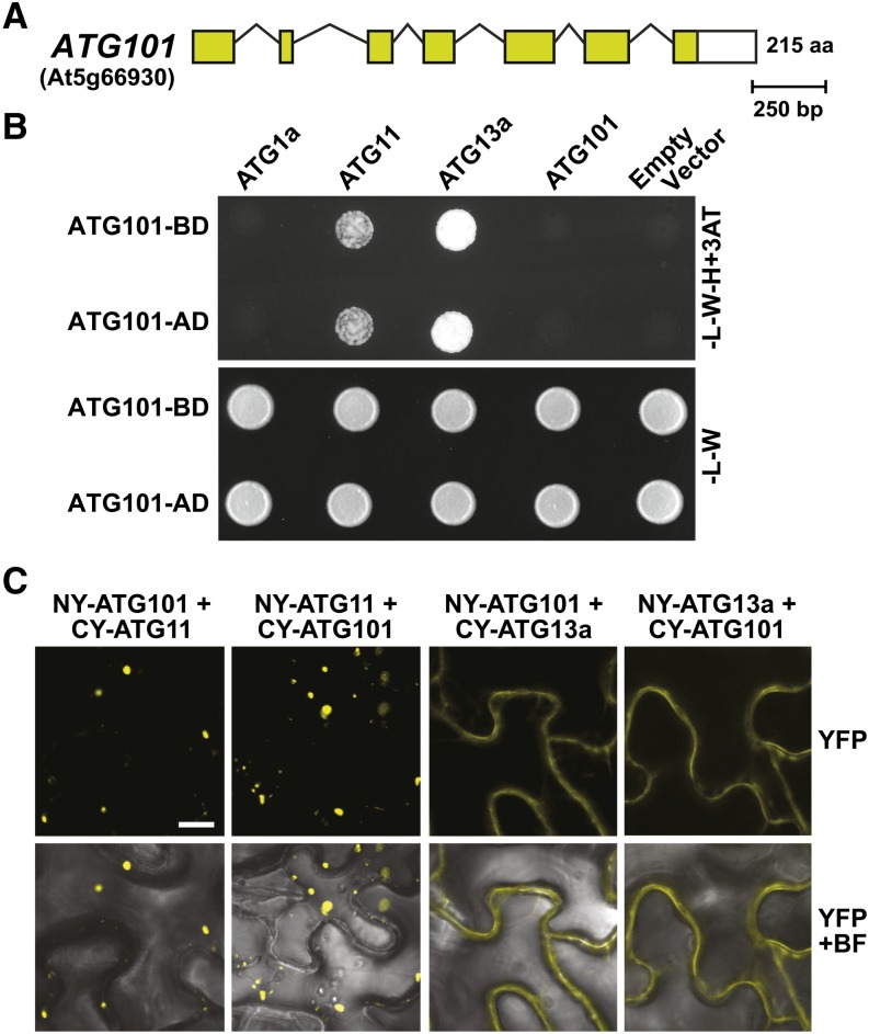
(A) Structure of the ATG101 locus (At5g66930). Lines represent introns, and the colored and white boxes represent coding and untranslated regions, respectively. Amino acid (aa) sequence length is indicated on the right.
(B) Y2H interactions of ATG101 with other subunits of the ATG1/13 complex. Full-length ATG101 designed as N-terminal fusions to either the GAL4 activating (AD) or binding (BD) domain was coexpressed with complementary AD or BD fusions of ATG1a, ATG11, and ATG13a. Shown are cells grown on selection medium lacking Trp and Leu (−L−W) or lacking Trp, Leu, and His and containing 3-amino-1,2,4-triazole (−L−W−H+3AT).
(C) Interaction of ATG101 with ATG11 or ATG13a in planta by BiFC. N. benthamiana leaf epidermal cells were coinfiltrated with plasmids expressing the N- and C-terminal fragments of YFP fused to ATG101, ATG11, and ATG13a. Reconstituted BiFC signals, as detected by confocal fluorescence microscopy of leaf epidermal cells 36 h after infiltration, are shown along with a bright-field (BF) image of the cells. Bar = 10 μm.

