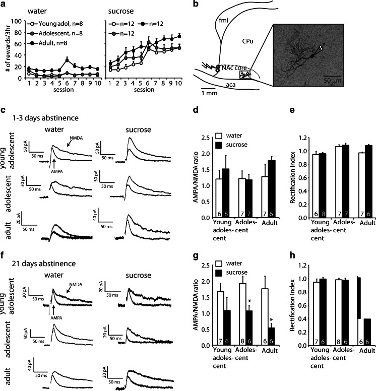
Fig. 3.
AMPA/NMDA ratios in the nucleus accumbens core after 1–3 days or 3 weeks of abstinence from sucrose or water self-administration. a Mean ± SEM number of earned rewards in rats trained to self-administer water (left) or sucrose (right) during the training phase. Brains were taken after 1–3 days or 21 days of abstinence without testing for cue-induced reward seeking. b Schematic of a parasagittal section through the nucleus accumbens core illustrating the relative placement of the stimulating electrode (bolt symbol) and recorded Neurobiotin-stained medium spiny neuron (inset) with respect to the forceps minor (fmi) and anterior commissure (aca). c Representative traces of medium spiny neurons held at +40 mV in the absence (both AMPA and NMDA) and presence (only AMPA) of the NMDAR antagonist AP5 in rats after 1–3 days of abstinence from self-administered water or sucrose at the three different age groups. Stimulation artifacts were removed for clarity. d Mean ± SEM AMPA/NMDA in NAc after 1–3 days of abstinence from self-administered water (white bars) or sucrose (black bars). The numbers indicate cells per group (n = 4–6 rats per group). No more than two cells per rat were used. e Rectification index after 1–3 abstinence days. f Representative traces of medium spiny neurons after 3 weeks of abstinence from water or sucrose self-administration in the three different age groups. g Mean ± SEM AMPA/NMDA ratio in NAc MSNs after 21 days of abstinence. The numbers indicate cells per group (n = 4–6 rats per group). No more than two cells per rat were used. h Rectification index after 21 abstinence days. *p < 0.05, different from the water condition within each age group

