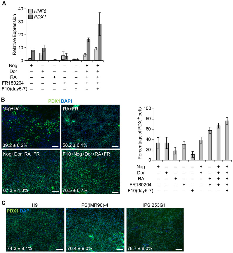
Figure 2. Differentiation of DE cells into pancreatic progenitor cells.
(A) Quantitative PCR analysis of the expression of HNF6 and PDX1 at day 10. Expression levels were normalized to TBP expression. mRNA expression was relative to that in control cells at day 10. Control cells were treated with three factors for 4 days as shown in Fig. 4A, and then without factors for 6 days. Error bars indicate SD (n = 3). (B) Left: cells were treated with each factor for 6 days and then stained with an anti-PDX1 antibody at day 10. Right: percentage of PDX1+ cells among differentiated cells treated with each factor. Mean ± SD (n = 5–10). (C) Each cell line, H9, iPS (IMR90)-4 and iPS 253G1, was treated with five factors for 6 days as shown in Fig. 4A, and stained then with an anti-PDX1 antibody at day 10. Mean ± SD (n = 5–10). F10, 50 ng/ml FGF10; Nog, 50 ng/ml Noggin; Dor, 1 μM dorsomorphin; RA, 2 μM retinoic acid; FR, 3 μM FR180204. Scale bar, 100 μm.

