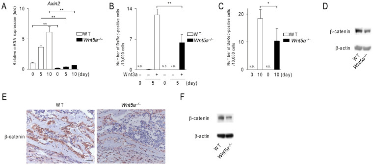
Figure 3. Wnt5a-deficient calvarial cells exhibit impaired Wnt/β-catenin signals.
(A) The expression of Axin2 in wild-type (WT) and Wnt5a−/− calvarial cell cultures. Cells were cultured in osteogenic medium for the indicated time and subjected to real-time PCR analysis. (B, C) The number of DsRed-positive cells in WT and Wnt5a−/− calvarial cell cultures transfected with the Tcf/Lef DsRed reporter adenovirus. In (B), cells were treated with Wnt3a (100 ng/ml) under growth culture conditions. In (C), cells were cultured under osteogenic conditions. (D) Western blot analysis of β-catenin expression in WT and Wnt5a−/− calvarial cells. Cells were cultured in osteogenic medium for 10 days. (E) Immunohistochemical staining (brown) of β-catenin in the scapulae of WT and Wnt5a−/− mice at E18.5. (F) Western blot analysis of β-catenin in the scapulae from WT and Wnt5a−/− mice at E18.5. Scale bar, 30 μm. In (A–C), data are expressed as the mean ± SD (n = 3–4). *p < 0.05, **p < 0.01, N.D.; not detected. In (D, F), the full length blots were presented in Supplementary Fig. S5.

