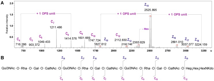
Figure 6.
(A) Part of the charge-deconvoluted ESI FT-ICR mass spectrum (negative ion mode) of the lower molecular mass fraction of the degraded PS isolated from the LPS of A. veronii Bs19, recorded with unspecific fragmentation. (B) Fragmentation scheme of the molecule. Mass numbers given refer to the monoisotopic masses. Mass fragments (marked with capital letters) are labeled according to the nomenclature of Domon and Costello [33].

