
Figure 4.
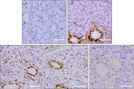
TUNEL staining in SMG. (a) Very few cells were stained in the control SMG. (b) One week after BTXA injection, numerous typical densely stained dark TUNEL-positive cell nuclei can be observed in ductal cells. (c) Two weeks after BTXA injection, apoptotic cells can be seen in the acinar and ductal cells. (d) TUNEL-positive cells decreased at 4 weeks after BTXA injection. (e) TUNEL-positive cells almost disappeared in the glands at 12 weeks after BTXA injection. Magnification ×400. Bar=50 μm. BTXA, botulinum toxin type A; SMG, submandibular gland; TUNEL, terminal deoxynucleotidyl transferase-mediated deoxyuridine triphosphate-biotin nick-end labeling.