Figure 3.
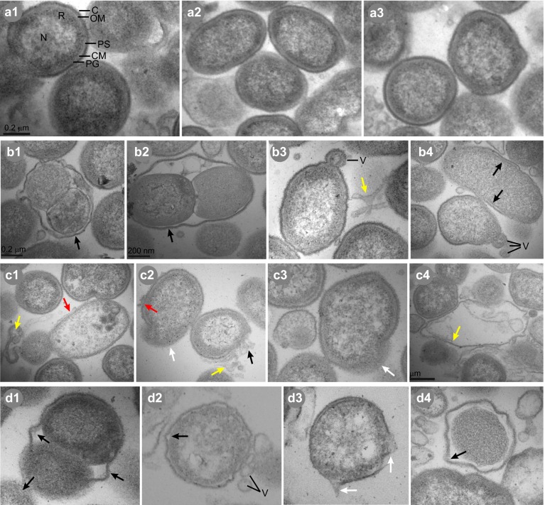
TEM images showing the ultrastructural effects of sphingoid bases and fatty acids on P. gingivalis. Untreated cells (a1–a3) exhibited typical Gram-negative coccobacillus morphology with outer membrane (OM), capsule (C), periplasmic space (PS), peptidoglycan (PG), cellular membrane (CM), distinct nucleoid (N) and ribosomal (R) regions and outer membrane vesicles (V). One hour treatments of P. gingivalis with phytosphingosine (b1–b4), sapienic acid (c1–c4) or SMAP28 (d1–d4) resulted in cellular distortion relative to untreated bacteria. Evidence of ultrastructural damage is indicated by the colored arrows: separation of the outer membrane from the cytoplasmic membrane (red); missing pieces of membrane (black); leakage of cytoplasmic contents (white); and detached membrane lying adjacent to cells (yellow). TEM, transmission electron microscopy.

