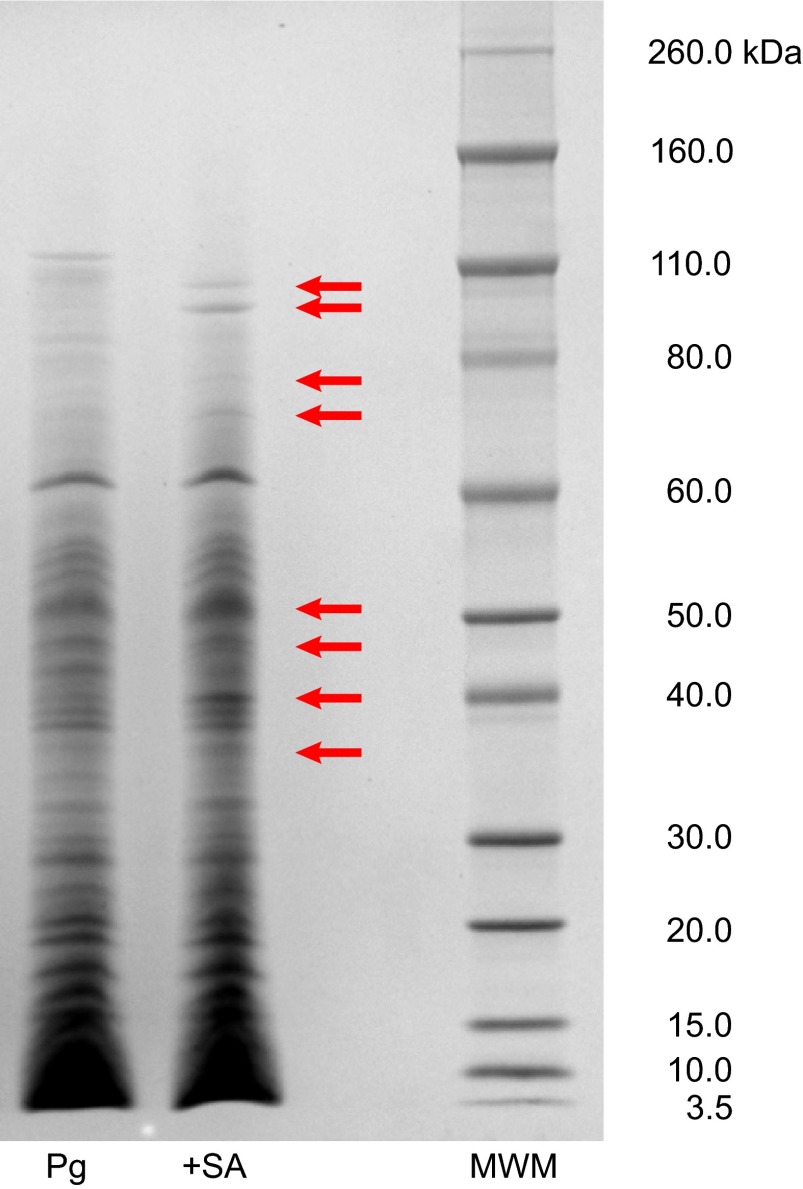
Figure 5.
SDS–PAGE separation of proteins in untreated and sapienic acid-treated P. gingivalis. Untreated (Pg) and sapienic acid-treated (+SA) proteins were separated by SDS–PAGE and visualized using Coomassie blue stain. SDS-PAGE, sodium dodecyl sulfate polyacrylamide gel electrophoresis; MWM, molecular weight marker, Novex sharp protein standards.

