
Figure 5. Truncated kinesins exhibit different patterns of accumulation early in neuronal development.


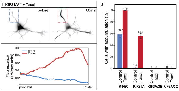
Truncated KIF1B (A) and KIF21B (D) accumulated at all neurite tips whereas truncated KIF17 (B) only accumulated at a subset of neurite tips. The remaining kinesins failed to accumulate at any tips. For KIF13-Fv constructs, cells were incubated with 10nM AP21087 for 4–6h before imaging. (I) KIF21A,, which is unable to accumulate in immature neurons, translocated to neurite tips following Taxol treatment (10 nM) for 1 h. Line scans of the average intensity along the neurite in the boxed region (below) confirm the redistribution of KIF21A417-GFP. (J) The accumulation of KIF21A and KIF5C but not Kinesin-2s in Stage 2 cells was enhanced by 10nM Taxol treatment for 4h. The figure shows the percentage of cells with truncated kinesin accumulation in at least one neurite; values are means and SEMs from 3 independent experiments. Dissociated hippocampal neurons were co-electroporated with tdTomato and a truncated kinesin or with a pair of truncated KIF3s. Scale bar: 20μm.