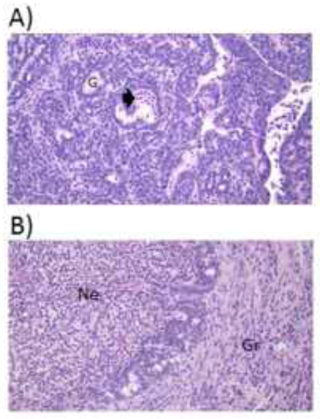
Figure 7.

Tumor inflammation and necrosis. Representative 5 μm sections stained with hematoxylin and eosin of (A) a small (0.3 g) and (B) a large (6.0 g) tumor. All tumors were pathologically similar: invasive ductal carcinomas with moderate differentiation. Smaller tumors exhibited minimal necrotic and inflammatory tissue, whereas larger tumors consisted of widespread necrosis with acute inflammation, as well as granulation tissue with chronic inflammation. Magnification = 400X. Arrowhead=acute inflammation and necrosis, G=gland, Ne=necrotic tissue with acute inflammation, Gr=granulation tissue with chronic inflammation
