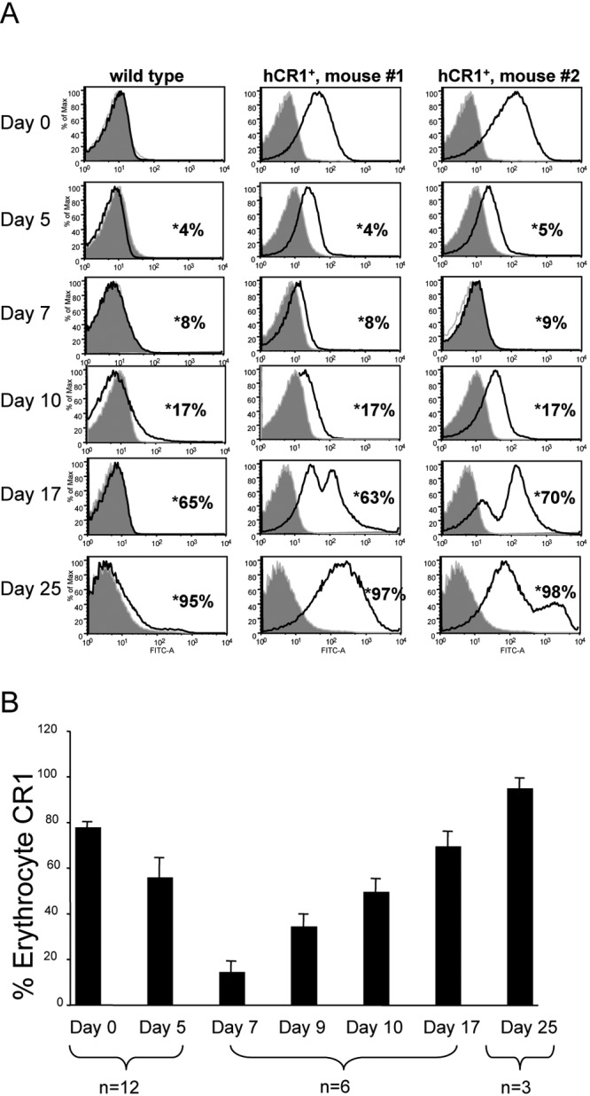
FIG 2 .

Infection of hCR1+ mice with P. berghei ANKA results in diminished detection of hCR1 on erythrocytes at 7 to 10 days postinfection. (A) Flow cytometry histograms from an experiment with one representative wild-type and two representative hCR1+ mice that survived 25 days following P. berghei ANKA infection are shown over a time course. Freshly isolated erythrocytes from infected or uninfected mice were analyzed on days 0, 5, 7, 9, 10, 17, and 25. The expression of cell surface hCR1 was analyzed. The histograms show the fluorescence intensity of erythrocyte CR1 detected (the x axis indicates FITC or CR1, and the y axis represents the percentage of the maximum); 1 × 10⁵ cells were analyzed in each event. Percent parasitemia of each mouse, also measured by flow cytometry (data not shown), is indicated with asterisks. (B) Average values of CR1 detection for each time point for available mice are shown. Error bars indicate the standard errors of the means.
