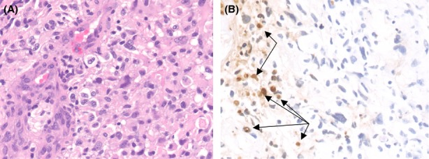
Figure 1.

Neuropathology results for 71-year-old male patient with high-grade glioma show increased cellularity, pleomorphic nuclei, and endothelial proliferation (A, original magnification 400×) with focal areas of necrosis characteristic of a GBM. Terminal deoxynucleotidyl transferase dUTP nick end labeling in situ hybridization (B) shows scattered positive nuclei within the tumor often associated with necrotic areas, however, other areas of the tumor not associated with necrosis also showed apoptosis (arrows, original magnification 400×).
