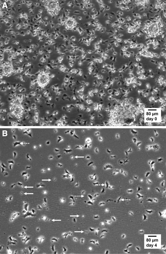
Fig. 2.

Still micrographs extracted from live cell time-lapse imaging of mussel hemocytes in culture. Hemocytes in suspension in hemolymph were collected, plated and cultured in MPSS at 14 °C as described in “Materials and methods” section and in Fig. 1. Time-lapse recordings were performed at 14 °C in a Peltier-cooling microscope stage with ×10 phase contrast objective lens. a Representative image selected from supplementary video_3 (duration 30 min, 1 image per 30 s), recorded in the droplet of hemolymph, immediately after the initial 20-min phase of adherence and before adding MPSS. Notice the high cell density and the presence of hemocyte aggregates. b Representative image selected from supplementary video_4 (duration 30 min, 1 image per 30 s), recorded after 4 days of culture in MPSS. Spread bipolar cells covered large distances by amoeboid movements (dotted arrows), while small round star-shaped hemocytes displayed static filopodia protrusions (solid arrows)
