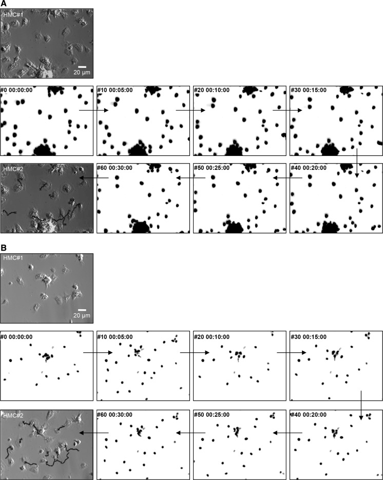
Fig. 3.
Determination of cell trajectories and velocities by live cell nuclei tracking. Hemocyte nuclei were specifically stained with Hoechst 33342. Wide field epifluorescence microscopy was used for time-lapse recording of nuclei movements under ×40 magnification. a and b correspond to distinct recordings (video_5 and video_6), where initial positions of hemocytes in the microscope field are shown in a first Hoffman modulation contrast micrograph (HMC#1). Several inverted images of hemocyte nuclei fluorescence at 5 min intervals are shown. A final HMC micrograph is presented at the end of the sequence (HMC#2) with different reconstituted nuclei trajectories superimposed

