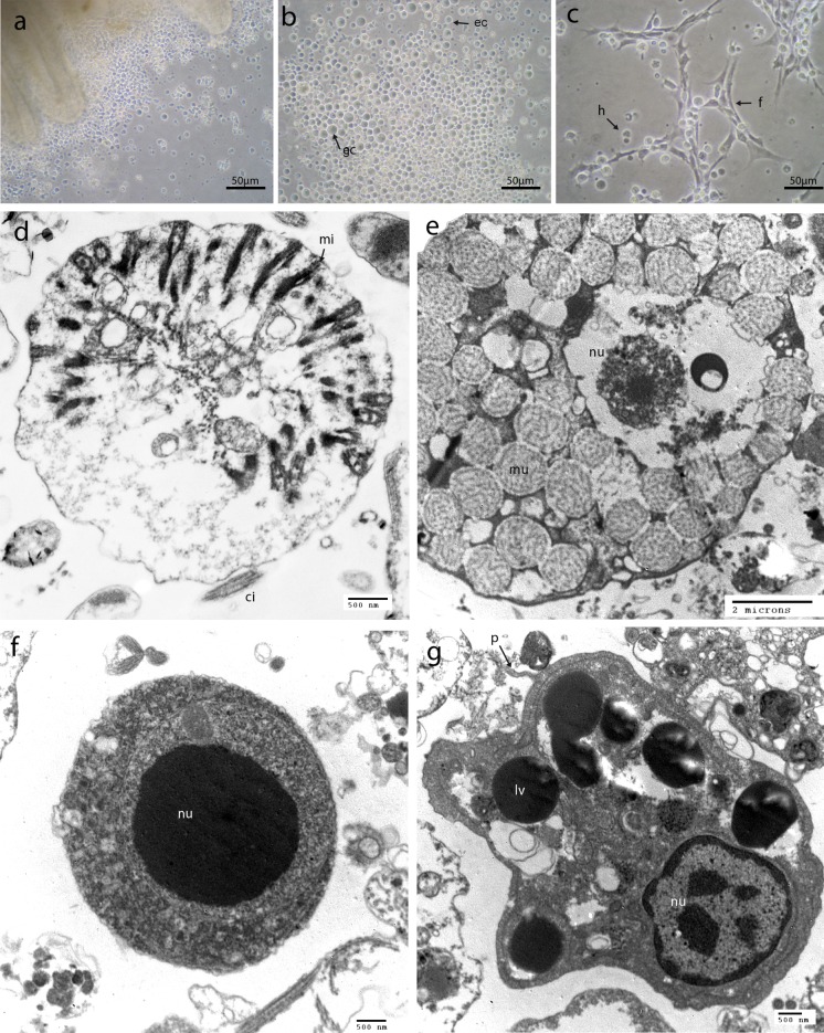
Fig. 1.
Abalone gill cell primary cultures observed under inverted phase contrast microscope (a, b, c) and transmission electron microscopy (d, e, f, g). a One-day-old gill explant primary gill cells culture from H. tuberculata showing the outgrowth of cells from an explant in a 6-well plate, b three day-old explant primary culture showing cell spreading around the flask bottom. Cell population consisted of rounded epithelial cells (ec), and glandular cells (gc) c three day-old explant culture showing hemocytes represented by small hyalinocytes (b) and fibroblastic like cells (f). Transmission electron micrograph of four-day-old cell culture d epithelial cell, e mucous cell, f hyalinocyte, g granulocyte. ci: cilia, nu: nucleus, mi: microvilli, mu: mucous vesicle, p: pseudopodia of the phagocytosis vesicle in formation, lv: lipofuschin vesicles

