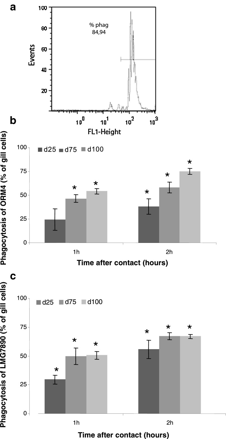
Fig. 3.
Phagocytosis by H. tuberculata gill cells estimated by flow cytometry. Phagocytosis index was expressed as percentage of gill cells containing three or more beads after 1 h of contact (a). Percentage of phagocytosis of gill cells in presence of the pathogenic ORM4 (b) and the non-pathogenic LMG7890 (c) bacterial strain after one and 2 h of contact with three densities of bacteria: 25, 75 and 100 bacteria/cell. Data are the mean ± SE of triplicate experiment. Significant differences (p < 0.05) from absorbances measured in control cells are indicated by an asterisk

